Diffuse Optical Tomography (DOT)
Near-infrared spectroscopy (NIRS) uses light to measure our brain activities. Due to its simplicity and non-invasive nature, it has been attracting attention as a way to extend brain measurement from the laboratory to daily environments.
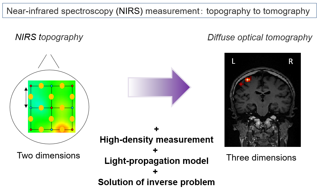
However, NIRS only provides the information of brain activity on the scalp, not in the brain. To reveal brain activities from the NIRS data, we are developing “Diffuse Optical Tomography (DOT),” a three-dimensional image reconstruction of brain activity.
Conventionally, NIRS topography has been widely used as a simple way to localize brain activity from NIRS data. However, this method has a major problem: Errors in source locations are induced by various factors such as non-uniformity of measurement sensitivity, scalp blood flow artifacts, and unknown partial optical path lengths.
DOT solves this problem by using high-density measurement (15-mm distance between probes) and two analysis processes (light-propagation simulation and image reconstruction). It has been reported that images reconstructed from NIRS data by DOT closely approximate the results of fMRIs (Eggbert et al., 2014).
In this laboratory, we aim to improve DOT by devising its algorithm for image reconstruction. We proposed a sparse DOT algorithm and showed that it improves the accuracy of depth and horizontal estimation over a conventional algorithm (depth-corrected least squares method) (Shimokawa et al., 2012).
Furthermore, we extended our method so that it could robustly reconstruct images even in the presence of scalp blood flow artifacts, as they exist in humans (Shimokawa et al., 2013).
Using real NIRS and fMRI data obtained during finger movements, we confirmed that the images reconstructed from the NIRS data by our method closely approximated the fMRI results (their differences were a few millimeters) (Yamashita et al., 2014).
In addition, using 12 subjects’ high-density NIRS data and fMRIs during three tasks, we showed that our method was robust against false positives (Yamashita et al., 2016). We are currently investigating its applicability to resting-state network analyses.
Collaborating with Richo Company, Ltd., we developed a multidirectional NIRS device, which can detect light from multiple directions, and showed that this device enables us to perform DOT even if the sensor arrays are not dense (Shimokawa et al., 2016).
References
- Eggebrecht, A. T., Ferradal, S. L., Robichaux-Viehoever, A., Hassanpour, M. S., Dehghani, H., Snyder, A. Z.,Culver, J. P. (2014). Mapping distributed brain function and networks with diffuse optical tomography. Nature Photonics, 8(6), 448–454
- Shimokawa, T., Ishii, T., Takahashi, Y., Sugawara, S., Sato, M., Yamashita, O., (2016), Diffuse optical tomography using multi-directional sources and detectors, Biomedical Optics Express, 7, No.7, pp.2623-2640
- Yamashita, O., Shimokawa, T., Aisu, R., Amita, T., Inoue, Y., Sato, M., (2016), Multi-subject and multi-task experimental validation of the hierarchical Bayesian diffuse optical tomography algorithm, NeuroImage, 135, 287-299
- Yamashita O, Shimokawa T, Kosaka T, Amita T, Inoue Y, and Sato M (2014), “Hierarchical Bayesian Model for Diffuse Optical Tomography of the Human Brain: Human Experimental Study”, Journal of Advanced Computational Intelligence and Intelligent Informatics, Vol.18, No.6 pp. 1026-1033
- T. Shimokawa, T. Kosaka, O. Yamashita, N. Hiroe, T. Amita, Y. Inoue, and M. Sato, “Extended hierarchical Bayesian diffuse optical tomography for removing scalp artifact,” Biomed. Opt. Express 4, 2411-2432 (2013)
- T. Shimokawa, T. Kosaka, O. Yamashita, N. Hiroe, T. Amita, Y. Inoue, and M. Sato, “Hierarchical Bayesian estimation improves depth accuracy and spatial resolution of diffuse optical tomography,” Opt. Express 20, 20427-20446 (2012)